Quick Guide Summary
In this comprehensive medical guide to Maxilla and Mandible fractures, we explore:
1. What are Maxillofacial Fractures?
Maxillofacial fractures refer to broken bones within the facial structure, primarily involving the Maxilla (the upper jaw, which houses the upper teeth and forms the floor of the nasal cavity) and the Mandible (the lower jawbone, which is a U-shaped bone holding the lower teeth and connecting to the skull via the temporomandibular joint, or TMJ).
These injuries most commonly result from high-impact trauma, such as road traffic accidents (RTAs), physical altercations, severe falls, or sports injuries. Because the jaws dictate your ability to chew, speak, and breathe comfortably, prompt surgical intervention is a critical medical necessity to restore proper alignment and physiological function.
2. Identifying a Jaw Fracture (Clinical Signs)
While severe swelling and bruising are obvious signs of trauma, specific clinical indicators point directly to a structural fracture of the maxilla or mandible:
Malocclusion
The most reliable sign of a jaw fracture. The patient feels that their teeth do not fit or bite together properly anymore, indicating the bone has shifted out of its natural alignment.
Paresthesia (Numbness)
Fractures of the mandible often compress or sever the inferior alveolar nerve, resulting in tingling or complete numbness in the lower lip and chin on the affected side.
Trismus (Restricted Opening)
Severe muscle spasms or structural blockage at the TMJ makes it extremely painful or physically impossible to open the mouth normally.
Intraoral Bleeding & Mobile Teeth
A "step" or gap may be felt along the gumline. Teeth near the fracture line may become highly mobile, or there may be persistent bleeding from the gum tissue where the bone has broken.
3. Maxilla vs. Mandible: Understanding the Break
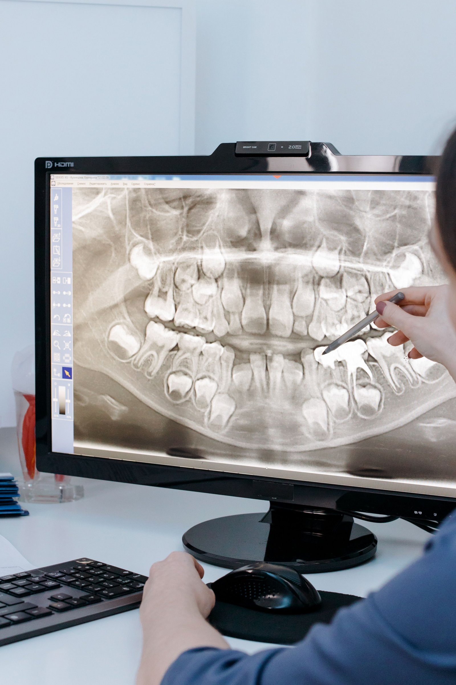
The surgical approach heavily depends on which bone is fractured and the specific anatomical zone of the break.
Maxillary Fractures (Le Fort Classifications)
The upper jaw is firmly attached to the skull. Fractures here are classified by René Le Fort's system based on the level of the break:
- • Le Fort I: A horizontal fracture right above the teeth roots (a floating palate).
- • Le Fort II: A pyramidal fracture extending upward to the nasal bridge.
- • Le Fort III: A transverse fracture that completely detaches the facial bones from the cranial base (craniofacial separation).
Mandibular Fractures
Because the lower jaw projects outward, it is the most frequently fractured facial bone. Common fracture zones include:
- • Condylar: Near the TMJ joint. Often caused by a direct blow to the chin.
- • Angle: At the back corner of the jaw, frequently complicated by impacted wisdom teeth creating a weak point in the bone.
- • Symphysis / Parasymphysis: Fractures occurring in the anterior (front) chin area.
4. The Surgical Treatment Protocol
Treating a jaw fracture follows orthopaedic principles: the bones must be realigned into their correct anatomical position (reduction) and held rigidly in place until they heal (fixation).
Phase 1: Emergency Stabilization & 3D Imaging
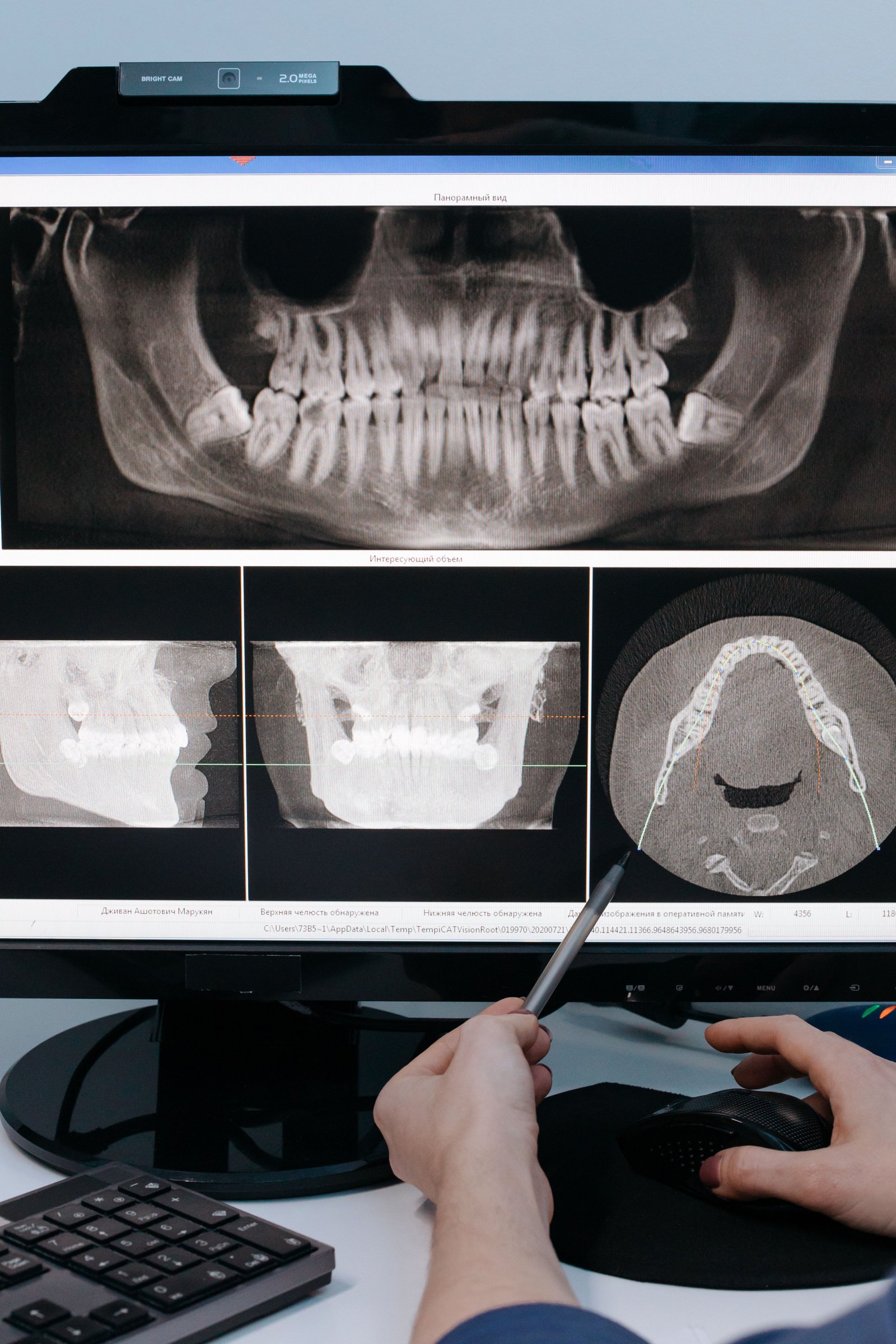
Airway management and halting hemorrhage are the first priorities in facial trauma. We then utilize advanced 3D CBCT (Cone Beam Computed Tomography) to map the exact, three-dimensional location of the fracture lines and any displaced bone fragments.
Phase 2: Reduction (Realigning the Bone)
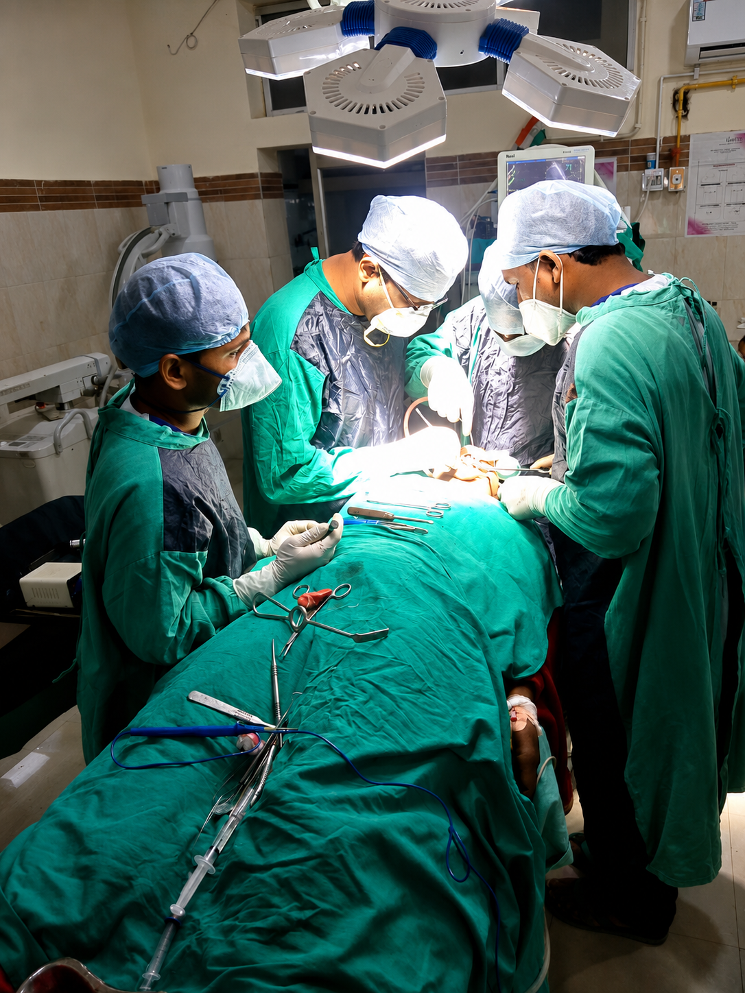
Under general anesthesia, the surgeon manually manipulates the broken jaw segments back into their proper anatomical positions. The teeth are used as a guide to ensure the original bite (occlusion) is perfectly restored.
Phase 3: Fixation (MMF vs. ORIF)
MMF (Maxillomandibular Fixation): The older, traditional method where the upper and lower teeth are wired together, literally wiring the jaw shut for 4 to 6 weeks to allow bone healing.
ORIF (Open Reduction & Internal Fixation): The modern gold standard. The surgeon makes an incision (often inside the mouth to hide scars) and uses specialized micro-titanium plates and screws to rigidly bolt the bone segments together. This often eliminates the need to wire the jaws shut.
Phase 4: Post-Operative Healing & Rehabilitation
The initial soft tissue heals in about 2 weeks, while the bone requires 6 to 8 weeks for clinical union. Strict dietary modifications and eventual physical therapy to restore normal jaw opening are mandatory.
Recovery & The Liquid Diet Protocol
Regardless of whether you receive MMF wiring or ORIF plating, the jawbone cannot endure chewing forces while it attempts to fuse. A strict non-chew diet is an absolute requirement for 4 to 6 weeks.
- • Weeks 1-2 (Pure Liquid): Protein shakes, blended soups, broths, and meal replacement drinks. No chewing whatsoever.
- • Weeks 3-6 (Soft Non-Chew): Mashed potatoes, scrambled eggs, yogurt, and very soft pastas that can be swallowed without grinding.
- • Oral Hygiene: If jaws are wired, standard brushing is impossible. You will use prescribed antimicrobial mouthwashes (Chlorhexidine) and a water flosser on low settings to prevent severe infection at the surgical sites.
5. Surgical Expertise in Varanasi
Managing severe facial trauma requires elite surgical proficiency and immediate access to advanced diagnostic imaging. Located strategically on BHU Trauma Centre Road in Lanka, Varanasi, Ayaansh Dental Clinic is ideally positioned to handle complex maxillofacial reconstructions.
The clinic is led by Dr. N Srivastava, a highly qualified specialist (MDS, BHU). Her extensive background in prosthodontics and implantology ensures that post-trauma jaw reconstruction not only stabilizes the bone but meticulously preserves the exact functional bite and facial aesthetics. For complex multi-trauma cases, we collaborate with leading maxillofacial surgeons to deliver comprehensive hospital-grade care.
6. Frequently Asked Questions
Not always. While traditional Maxillomandibular Fixation (MMF) involves wiring the jaws together for 4 to 6 weeks, modern Open Reduction and Internal Fixation (ORIF) uses titanium plates and screws to stabilize the bone internally. This often allows patients to open their mouths and speak almost immediately after surgery, though a soft diet is still strictly required.
The initial soft tissue healing (gums and incisions) takes about 2 weeks. However, the jawbone typically requires 6 to 8 weeks to achieve primary clinical union, meaning it is stable enough for normal chewing. Complete microscopic bony remodeling can take several months.
In the vast majority of cases, no. Medical-grade titanium is highly biocompatible and can remain permanently in your face without causing issues. Plates and screws are only removed if they become infected, cause persistent cold sensitivity, or physically interfere with a future dental implant placement.
Immediately after surgery, you will experience significant swelling and bruising, which alters your appearance temporarily. However, the primary surgical goal of reduction is to realign the bones exactly as they were before the trauma. Once fully healed, your facial profile and bite should return to their normal, pre-injury state.

Dr. N Srivastava
AuthorBDS, MDS (BHU) • Prosthodontist & Implantologist
An expert trained at the prestigious Banaras Hindu University (BHU), Dr. N Srivastava specializes in complex dental implant surgery, full mouth reconstructive prosthodontics, and oral surgical procedures. She utilizes advanced 3D diagnostics and stringent surgical protocols to deliver optimal functional recovery for patients in Varanasi.