Quick Guide Summary
In this highly specialized guide to Ocular Prosthetics, we explore:
1. What is an Ocular Prosthesis?
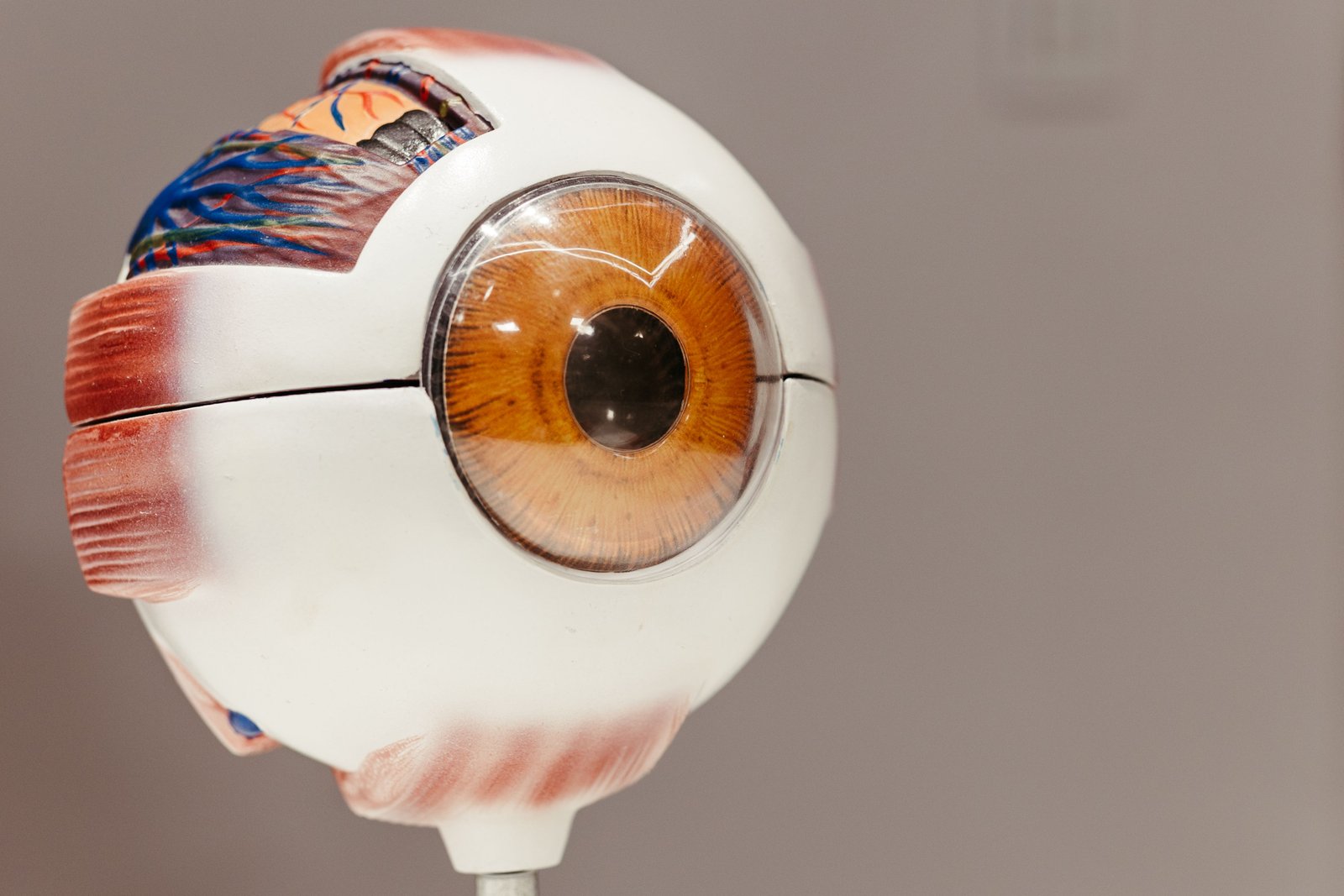
An Ocular Prosthesis, commonly referred to as an artificial eye, is a highly customized cranial prosthesis designed to replace an absent natural eye. It falls under the extremely specialized dental sub-field of Maxillofacial Prosthodontics.
It is important to understand that an artificial eye cannot restore vision. Its purpose is entirely structural and cosmetic. It fits directly over an orbital implant (placed by an ophthalmologic surgeon) under the eyelids. Crafted from high-grade, biocompatible PMMA (acrylic resin), the prosthesis restores the natural volume of the eye socket, prevents the eyelids from collapsing, allows for proper tear drainage, and perfectly mirrors the appearance of the remaining healthy eye.
2. Clinical Indications (Why is it needed?)
The loss or absence of an eye is a profound psychological and physical trauma. Prosthetic intervention is medically indicated in several primary scenarios:
Oncology (Eye Cancer)
Malignant intraocular tumors, such as Retinoblastoma in children or Uveal Melanoma in adults, often require the complete removal of the eyeball to prevent the cancer from spreading to the brain or the rest of the body.
Severe Trauma
Irreparable globe ruptures or penetrating injuries resulting from road traffic accidents, industrial accidents, or sports injuries where the eye's architecture cannot be surgically salvaged.
Congenital Defects
Conditions like Anophthalmia (born entirely without an eye) or Microphthalmia / Phthisis bulbi (a severely shrunken, non-functional eye). Early prosthetic intervention in children is vital to stimulate normal facial bone growth.
Painful Blind Eye
End-stage conditions like severe glaucoma or chronic untreatable infections can result in an eye that is completely blind but causes relentless, excruciating pain, necessitating its removal.
3. Surgical Prerequisites
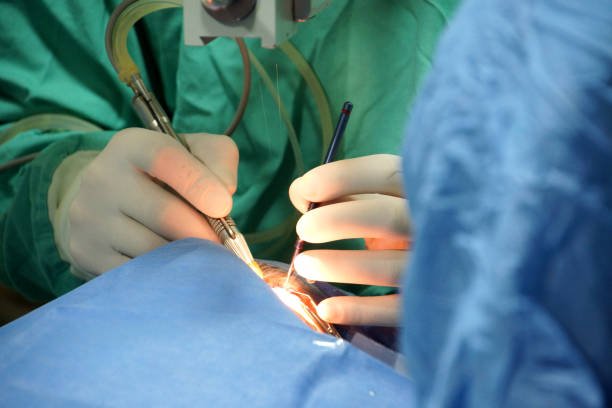
Before the prosthodontist can craft the artificial eye, an ophthalmologist or oculoplastic surgeon must perform the removal surgery. The type of surgery dictates how the final prosthesis will fit and move.
Evisceration
The surgeon removes the inner jelly-like contents of the eye (cornea and intraocular contents) but leaves the outer white shell (sclera) and the attached eye muscles entirely intact. A spherical orbital implant is placed inside the scleral shell. Result: Because the natural muscles remain perfectly attached, an ocular prosthesis fitted over an evisceration generally has the most natural, synchronous movement.
Enucleation
The complete removal of the entire eyeball, severing it from the optic nerve and the eye muscles. This is standard protocol for ocular tumors. The surgeon places an orbital implant deep in the socket and surgically re-attaches the eye muscles to the implant itself. Result: Provides good movement, though slightly less dynamic than evisceration.
Exenteration
The most aggressive surgery, involving the removal of the eyeball, the eyelids, and all surrounding orbital tissues down to the bone. Result: The patient requires a much larger "Orbital Prosthesis" (which includes artificial eyelids and silicone skin) rather than a simple ocular shell.
4. The Artistic & Clinical Workflow
Fabricating an ocular prosthesis is an intricate blend of medical science and fine-art painting. The process requires several meticulous appointments, usually beginning 6 to 8 weeks post-surgery, once the swelling has completely subsided.
Socket Impression
We inject a gentle, specialized medical silicone impression material directly into the healed eye socket. This captures the exact, unique contours of the tissue bed and the orbital implant, ensuring the final acrylic piece will fit like a perfect puzzle piece without irritating the tissue.
Wax Try-In & Sclera Fabrication
A wax model of the eye is created from the mold. The patient tries this on, and the prosthodontist meticulously carves the wax to ensure the eyelids drape naturally and perfectly match the opposite side. The wax is then converted into a white acrylic "sclera" (the white of the eye).
Hand-Painting the Iris & Veins
This is the most critical artistic step. With the patient present in the clinic, the specialist hand-paints the iris using specialized pigments to perfectly replicate the exact color, stroma patterns, and pupillary size of the contralateral healthy eye. Tiny red silk threads are applied to the white acrylic to mimic vascularization (blood veins).
Final Curing & High-Shine Polishing
A clear acrylic cap is processed over the painted details to lock them in permanently. The entire prosthesis undergoes rigorous, multi-stage polishing. The surface must be as smooth as glass so the inner eyelids glide over it effortlessly and natural tear fluid spreads evenly.
Care, Maintenance, and Lifespan
Unlike a dental denture, an ocular prosthesis is not removed daily. Excessive handling can introduce bacteria into the socket and stretch the delicate eyelid tissues.
- • Routine Cleaning: Most patients only remove their artificial eye once every 1 to 3 months for a gentle wash with mild, unscented soap and water. Removal and insertion are done easily using a specialized rubber suction cup.
- • Professional Polishing: Protein and salt deposits from your tears will slowly accumulate on the acrylic surface, causing irritation. You must visit the clinic once a year for a professional high-speed polish.
- • The Lifespan: Tissues in the eye socket naturally shift and settle with age, and acrylic slowly degrades. A typical custom ocular prosthesis needs to be replaced every 5 to 7 years for adults, and more frequently for growing children.
6. Maxillofacial Expertise in Varanasi
Fabricating an undetectable ocular prosthesis is an art form mastered by very few dental professionals. Located strategically on BHU Trauma Centre Road in Lanka, Varanasi (easily accessible from Samneghat, Assi Ghat, and Ravidas Gate), Ayaansh Dental Clinic offers this highly specialized, life-changing service.
The clinic is directed by Dr. N Srivastava, a highly qualified MDS Prosthodontist. Her advanced clinical training at Banaras Hindu University (BHU) equipped her with the exact anatomical and artistic skills required to rehabilitate patients suffering from severe facial and ocular defects, working in tandem with the region's leading ophthalmologists.
7. Frequently Asked Questions
No. An ocular prosthesis is purely cosmetic and structural. It cannot restore sight, but it restores the natural volume of the eye socket, supports the eyelids, and harmonizes facial symmetry.
Yes, to a certain extent. If the surgeon placed an orbital implant connected to your natural eye muscles during the removal surgery, the prosthesis will sit flush against this implant. When your muscles move the internal implant, the visible acrylic prosthesis will track and move synchronously with your healthy eye.
No. The eye socket is completely healed and desensitized before the prosthesis is placed. Once properly fitted and highly polished, you will not even feel the prosthesis under your eyelids.
Yes. In fact, it is highly recommended to leave the ocular prosthesis in 24/7. Removing it constantly can cause the socket tissues to shrink and irritate the delicate linings of the eyelids.
Historically, they were known as "glass eyes," but today, glass is rarely used. Modern ocular prostheses are fabricated from extremely tough, medical-grade PMMA (acrylic resin), which is shatterproof, highly adaptable, and much safer for the patient.

Dr. N Srivastava
AuthorBDS, MDS (BHU) • Prosthodontist & Implantologist
An expert trained at the prestigious Banaras Hindu University (BHU), Dr. N Srivastava specializes in complex dental implant surgery, full mouth reconstructive prosthodontics, and highly specialized maxillofacial rehabilitation. She utilizes advanced diagnostics and artistic precision to restore facial aesthetics and identity for trauma and oncology patients in Varanasi.