Quick Guide Summary
In this comprehensive medical guide to Premalignant Lesions, we explore:
1. What is Leukoplakia?
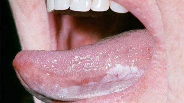
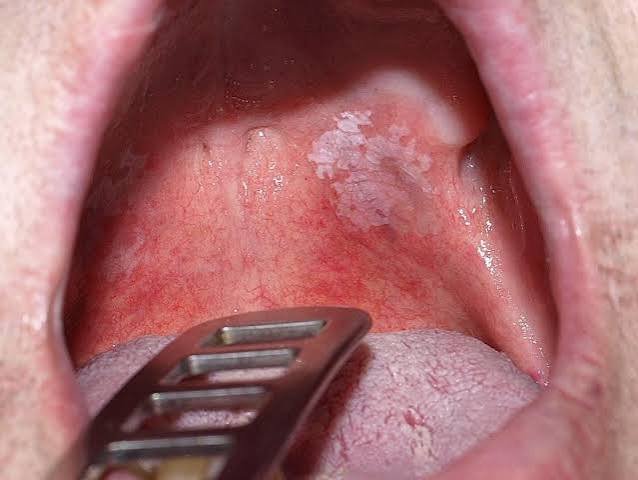
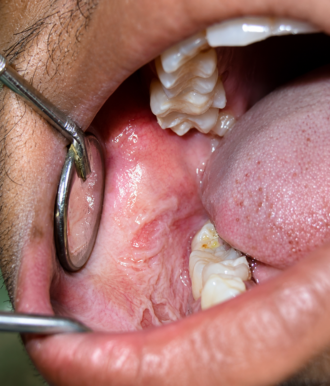
The term Leukoplakia originates from Greek, meaning "white patch." In clinical oral pathology, it is strictly defined as a predominantly white plaque or patch attached to the oral mucosa that cannot be rubbed or scraped off and cannot be characterized clinically or pathologically as any other defining disease.
These patches typically appear on the inside of the cheeks (buccal mucosa), the bottom of the mouth, or the borders of the tongue. While the patch itself is not cancer, it is classified by the World Health Organization (WHO) as a Potentially Malignant Disorder (PMD). This means that the cellular structure within the patch is abnormal and carries a statistically significant risk of transforming into Oral Squamous Cell Carcinoma (OSCC) over time.
2. Primary Causes and Risk Factors
Leukoplakia is the mouth's biological response to chronic irritation. When the delicate oral mucosa is constantly assaulted, it responds by rapidly producing an excess of keratin (hyperkeratosis) to protect itself, resulting in the thick white patch. The primary culprits include:
Tobacco & Areca Nut
By far the leading cause globally. Combustible tobacco (smoking) and smokeless tobacco (chewing tobacco, gutkha, betel nut, pan masala) constantly bathe the tissues in powerful chemical carcinogens.
Chronic Alcohol Consumption
Heavy alcohol use dehydrates the oral mucosa and acts as a solvent, making it significantly easier for tobacco carcinogens to penetrate deep into the cellular layers.
Mechanical Trauma
A jagged, broken tooth, an ill-fitting denture, or chronic cheek-biting can physically traumatize the tissue over months or years, leading to frictional hyperkeratosis.
Viral Infections (HPV)
Certain strains of the Human Papillomavirus (HPV) are increasingly being linked to cellular changes in the oral cavity that can present as premalignant lesions.
3. Clinical Diagnosis: The Role of the Biopsy
It is impossible for any doctor to determine if a white patch is cancerous merely by looking at it. While clinical experience guides suspicion, a biopsy is the absolute gold standard and mandatory requirement for a definitive diagnosis.
Incisional / Excisional Scalpel Biopsy
Under local anesthesia, a tiny, precise sample of the white patch (including a margin of normal tissue) is surgically removed. It is sent to an oral pathologist who examines the cellular architecture under a microscope. This is the only way to detect the presence of cancer cells.
Oral Brush Biopsy
A less invasive, preliminary screening tool where a specialized stiff brush is rotated over the lesion to collect transepithelial cells. While useful for initial screening, if atypical cells are found, a traditional scalpel biopsy must still follow.
4. Understanding Dysplasia & Malignant Risk
When the pathologist examines the biopsy, they are looking for Dysplasia. Dysplasia refers to abnormal cellular growth and maturation. It is the direct precursor to cancer. The severity of dysplasia dictates the immediate treatment plan.
Mild to Moderate Dysplasia
The cellular changes are present but confined to the lower layers of the tissue. If the underlying cause (like smoking) is eliminated immediately, these lesions can sometimes regress or remain stable. Strict, frequent clinical monitoring is required.
Severe Dysplasia / Carcinoma In Situ
The abnormal cells span the entire thickness of the tissue covering but have not yet broken through the basement membrane into the deeper tissues. This is highly volatile and requires immediate, aggressive surgical removal to prevent it from becoming invasive oral cancer.
Erythroleukoplakia (The Red Flag)
If a leukoplakia patch develops speckled red areas within the white plaque (known as Erythroleukoplakia), the risk of malignant transformation skyrockets. Red patches indicate that the tissue is so abnormal that blood vessels are exposed. These must be biopsied with absolute urgency.
5. Modern Treatment Protocols
Treatment is entirely dependent on the biopsy results.
1. Risk Factor Elimination
The first and most crucial step. The patient must completely halt all tobacco and alcohol use. If a sharp tooth is the cause, it must be smoothed or extracted. In many cases of mild dysplasia, removing the irritant allows the patch to heal naturally.
2. Surgical Excision
For severe dysplasia or lesions that do not resolve after habits are stopped, the patch must be physically removed. This is done via a minor surgical procedure using a scalpel, ensuring clear, healthy margins.
3. Laser Ablation
Advanced medical lasers (like CO2 or Diode lasers) are increasingly used to vaporize the abnormal tissue. Lasers offer excellent precision, minimal bleeding, and often faster, less painful recovery times.
4. Lifelong Monitoring
Even after successful surgical removal, leukoplakia has a high recurrence rate. Patients require strict, lifelong follow-up appointments every 3 to 6 months to catch any new cellular changes immediately.
6. Specialized Oral Care in Varanasi
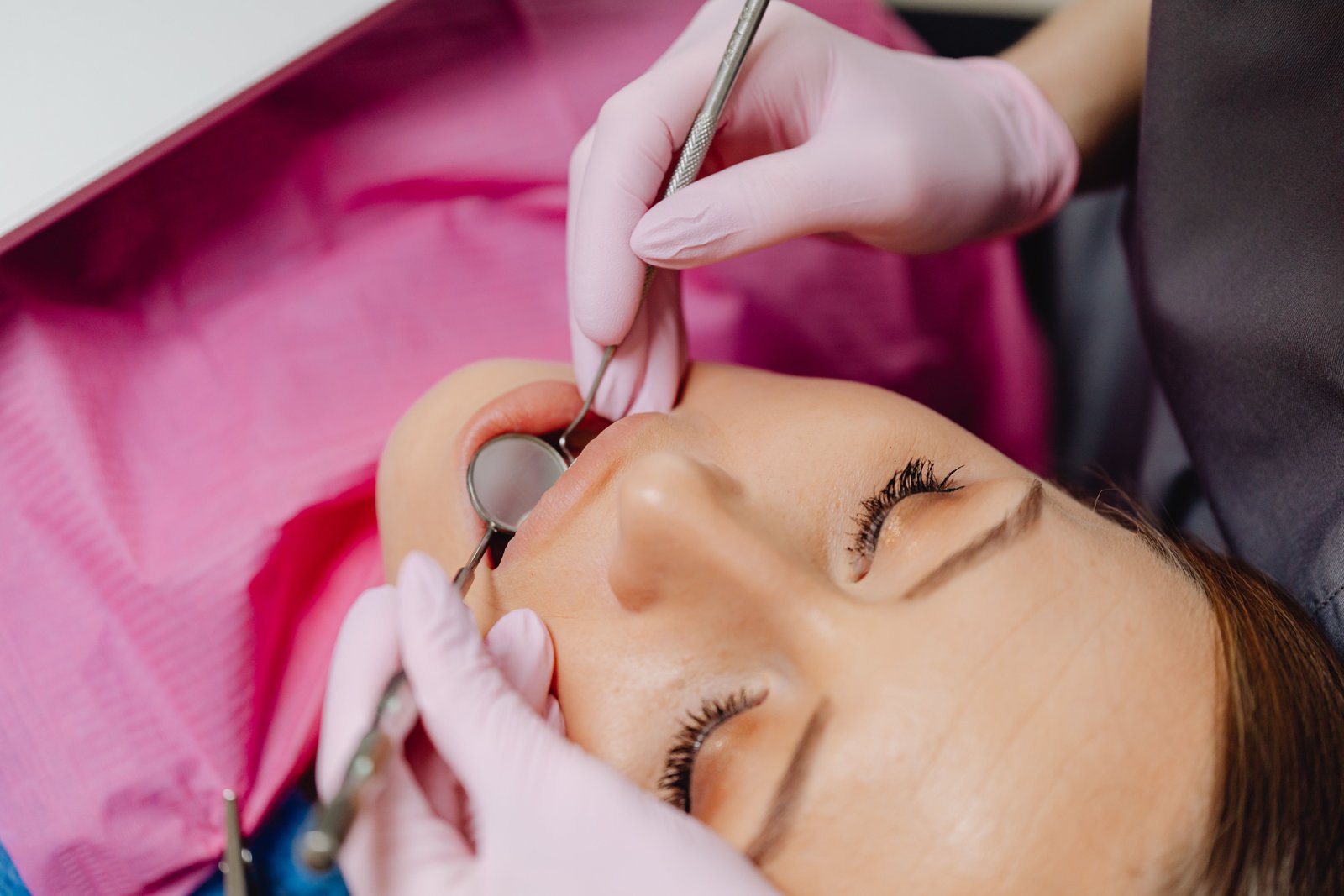
Detecting and managing premalignant lesions requires a highly trained clinical eye. Located strategically on BHU Trauma Centre Road in Lanka, Varanasi (easily accessible from Assi Ghat, Samneghat, and Ravidas Gate), Ayaansh Dental Clinic offers expert oral pathology screening.
Directed by Dr. N Srivastava, a specialist from Banaras Hindu University (BHU), our clinic emphasizes early detection. If a lesion is deemed high-risk or confirmed malignant, we work directly in tandem with the region's leading surgical oncologists and pathologists to ensure seamless, life-saving care and subsequent maxillofacial rehabilitation.
7. Frequently Asked Questions
Not necessarily. Leukoplakia is a 'premalignant' or potentially malignant lesion. While it is not cancer yet, it indicates that the cells are behaving abnormally and carry a significantly higher risk of turning into oral squamous cell carcinoma if left untreated.
Usually, it is completely painless, which makes it dangerous as patients often ignore it for months or years. However, if the patch becomes ulcerated, red, or hard, it may become sensitive to spicy or hot foods.
Yes, but only if it is a mild frictional or reactive leukoplakia and the irritant (like smoking, chewing tobacco, or a sharp tooth) is completely and permanently removed. If the patch does not resolve within 2 to 4 weeks after stopping the habit, a biopsy is mandatory.
Oral thrush is a fungal infection (Candida) that also presents as white patches in the mouth. The crucial diagnostic difference is that thrush patches can be easily scraped or wiped off with a gauze, revealing a red, raw surface underneath. Leukoplakia is firmly attached and cannot be scraped away.
Dr. N Srivastava
AuthorBDS, MDS (BHU) • Prosthodontist & Implantologist
An expert trained at the prestigious Banaras Hindu University (BHU), Dr. N Srivastava specializes in advanced diagnostics, full mouth rehabilitations, and complex oral restorations. She emphasizes early detection of oral pathologies, ensuring patients in Varanasi receive prompt, life-saving interventions.