Quick Guide Summary
In this comprehensive medical guide to Premalignant Lesions, we explore:
1. What are White and Red Lesions?
The oral cavity is lined by a delicate, pink mucous membrane. When this tissue is chronically irritated or begins to undergo abnormal cellular changes, it alters the visual clinical presentation. These alterations primarily manifest as distinct color changes—most notably white and red patches.
The World Health Organization (WHO) classifies these persistent patches as Potentially Malignant Disorders (PMDs). This implies that while the patch itself may not be cancerous at the exact moment of discovery, the cells within it are behaving erratically and carry a highly significant statistical risk of transforming into Oral Squamous Cell Carcinoma (OSCC) if left unmonitored and untreated.
2. Erythroplakia vs. Leukoplakia (The Danger of Red)
Differentiating between the color profiles of a lesion provides vital clues to the clinician regarding the severity of the cellular damage.
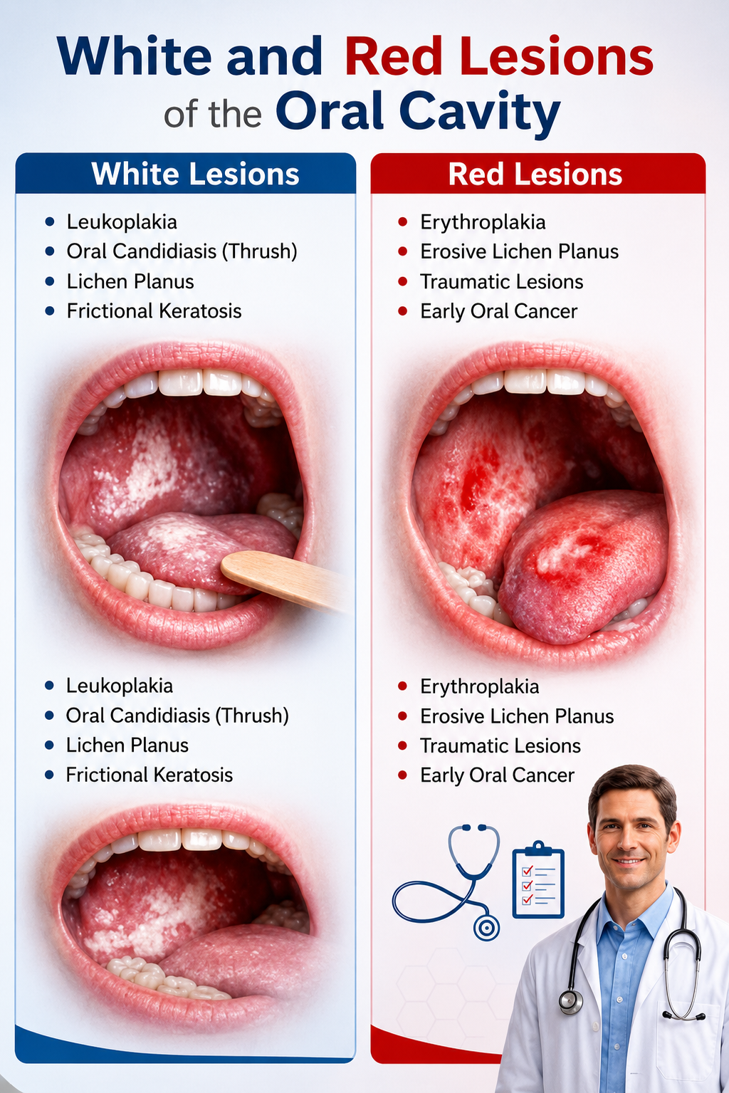
Leukoplakia (The White Patch)
Clinically defined as a predominantly white plaque that cannot be scraped off. The white color occurs because the tissue reacts to chronic irritation by rapidly overproducing keratin (hyperkeratosis) to build a protective callus. While concerning, purely homogenous white patches generally have a lower immediate risk of harboring severe dysplasia compared to red patches.
Erythroplakia (The Red Patch)
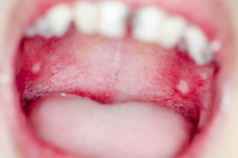
A fiery red, often velvety or granular patch that cannot be clinically attributed to inflammation from trauma or infection. The red color is a massive warning sign; it indicates that the protective outer layer of skin (epithelium) has thinned out so severely (atrophy) that the underlying blood vessels are visible. Erythroplakia has the highest risk of malignant transformation. Biopsies of true erythroplakia reveal severe dysplasia or carcinoma in situ in over 90% of cases.
Erythroleukoplakia (The Mixed Patch)
A highly volatile lesion presenting as a white patch scattered with distinct red, speckled, or nodular areas. The presence of red specks within a white plaque drastically elevates the risk profile, making immediate biopsy mandatory.
3. Primary Causes and Triggers
Premalignant lesions do not appear randomly. They are the oral mucosa's biological reaction to sustained chemical or mechanical damage over long periods.
Tobacco & Gutkha
Combustible smoking and smokeless tobacco (betel nut, pan masala, khaini) are the absolute leading culprits globally. They continuously bathe the oral tissues in hundreds of known, highly reactive carcinogens.
Heavy Alcohol Use
Alcohol acts as a solvent. It dehydrates the oral skin and breaks down lipid barriers, making it exponentially easier for tobacco toxins to penetrate deep into the vulnerable cellular layers.
Chronic Mechanical Trauma
A persistently sharp, broken tooth, an ill-fitting abrasive denture, or the nervous habit of chronic cheek-biting physically traumatizes the tissue, leading to thick, reactive white calluses.
Human Papillomavirus (HPV)
Certain high-risk strains of HPV (particularly HPV-16 and 18) are increasingly documented in causing specific cellular dysplasias in the oral cavity and oropharynx that manifest as distinct clinical lesions.
4. Diagnostic Protocol: The Role of the Biopsy
It is clinically impossible for any specialist to declare a white or red patch as benign or cancerous simply by visual inspection. A definitive diagnosis requires laboratory histopathology.
Vital Staining (Toluidine Blue)
An initial clinical adjunct. The dentist applies a specialized blue dye to the lesion. Dysplastic (abnormal) cells retain the blue dye much stronger than healthy cells, helping the surgeon identify the most volatile area to target for the physical biopsy.
Incisional Scalpel Biopsy (The Gold Standard)
Under profound local anesthesia, a small, precise segment of the patch is surgically removed, ensuring a margin of adjacent healthy tissue is included. The tissue is sent to an oral pathologist who examines the cellular architecture under a microscope to confirm or rule out the presence of Dysplasia.
5. Modern Management & Treatment
Treatment protocols are entirely dictated by the severity of dysplasia discovered in the biopsy report.
1. Risk Factor Eradication
The first and most critical intervention. The patient must completely halt all tobacco, gutkha, and alcohol use. In many cases of purely frictional, mild leukoplakia, removing the irritant allows the patch to regress naturally over several weeks.
2. Surgical Excision
For lesions exhibiting severe dysplasia, or high-risk red patches (erythroplakia), the entire lesion must be physically cut out. This minor surgical procedure ensures clear, healthy margins, preventing the abnormal cells from multiplying and invading deeper tissues.
3. Laser Ablation
Advanced medical lasers (like CO2 lasers) are highly effective in vaporizing large, widespread premalignant patches. Lasers offer extreme precision, instantly cauterize blood vessels for minimal bleeding, and generally result in a less painful, faster healing process.
4. Lifelong Monitoring
Oral mucosal diseases possess a high recurrence rate, a phenomenon known as 'field cancerization'. Even after successful surgical removal, patients must commit to strict follow-up appointments every 3 to 6 months for the rest of their lives.

6. Specialized Oral Care in Varanasi
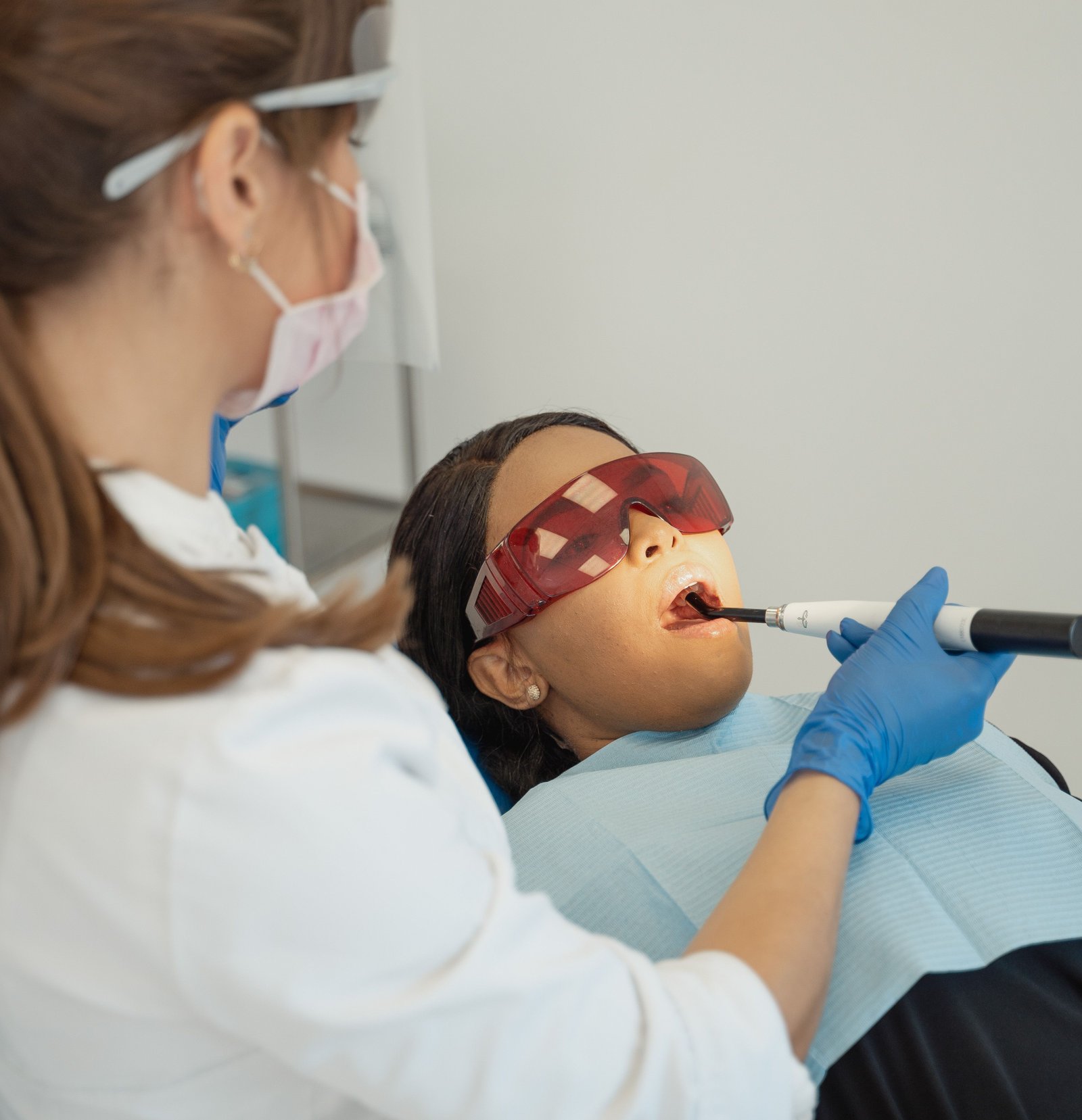
Detecting the subtle visual cues of a premalignant lesion requires a highly trained, specialized clinical eye. Located strategically on BHU Trauma Centre Road in Lanka, Varanasi (easily accessible from Assi Ghat, Samneghat, and Ravidas Gate), Ayaansh Dental Clinic offers expert oral pathology screening.
Directed by Dr. N Srivastava, a specialist from Banaras Hindu University (BHU), our clinic emphasizes aggressive early detection. If a lesion is deemed high-risk or confirmed malignant, we coordinate seamlessly with the region's leading surgical oncologists to ensure prompt, life-saving interventions and subsequent maxillofacial rehabilitation.
7. Frequently Asked Questions
Yes. While both require immediate medical attention, purely red patches (Erythroplakia) or mixed red-and-white patches carry a significantly higher risk of demonstrating severe dysplasia or invasive cancer upon biopsy compared to purely white, homogenous patches.
Purely white patches (Leukoplakia) are typically completely painless, which makes them dangerous as patients often ignore them. However, red lesions indicate a severe thinning of the protective oral skin, leaving nerves exposed. Patients with red lesions often report a burning sensation, especially when consuming hot or spicy foods.
An incisional biopsy is a very quick, minor surgical procedure. Conducted under profound local anesthesia right in the dental chair, the physical sampling takes only 10 to 15 minutes. A couple of dissolvable stitches are placed, and you can usually return to work the same day.
Yes. Chronic cheek biting or a persistently sharp tooth can cause a reactive white line or patch called "frictional keratosis" (essentially a callus). If the sharp tooth is smoothed and the patch disappears after two weeks, it was benign. If it persists, it must be biopsied.

Dr. N Srivastava
AuthorBDS, MDS (BHU) • Prosthodontist & Implantologist
An expert trained at the prestigious Banaras Hindu University (BHU), Dr. N Srivastava specializes in advanced diagnostics, full mouth rehabilitations, and complex oral restorations. She emphasizes early detection of oral pathologies, ensuring patients in Varanasi receive prompt, life-saving interventions.