Quick Guide Summary
In this comprehensive guide to dental imaging and diagnostics, we explore:
1. The Invisible World: Why We Need X-Rays
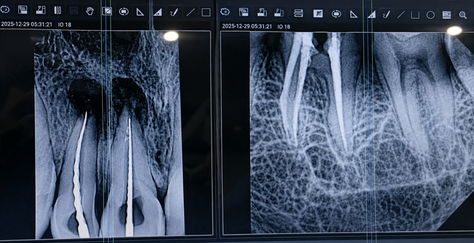
When a dentist performs a visual examination of your mouth using a mirror and a probe, they are only seeing about 30% of your actual dental structure. The remaining 70%—including the roots of the teeth, the surrounding alveolar jawbone, the ligaments, and the internal nerve chambers—is completely hidden beneath the gumline.
Without radiographic imaging (X-rays), it is biologically impossible to accurately diagnose early-stage interproximal decay (cavities forming tightly between two teeth), bone loss caused by periodontal disease, deep periapical abscesses, or impacted wisdom teeth. Attempting complex dental treatments without X-rays is akin to a surgeon operating blindfolded.
2. Modern Digital X-Rays: RVG and OPG
The era of biting down on uncomfortable cardboard films and waiting 15 minutes for them to develop in toxic darkroom chemicals is over. Dentistry has transitioned entirely to high-speed digital imaging.
RVG (Radiovisiography)
This is the modern equivalent of the small, localized X-ray. A tiny, highly sensitive digital sensor is placed inside the mouth. It instantly transmits a high-resolution, manipulatable image directly to a computer screen in real-time. It is used to closely examine 1 to 3 specific teeth for deep cavities, root canal anatomy, or local bone levels. RVG sensors require up to 80% less radiation than traditional film.
OPG (Orthopantomogram)
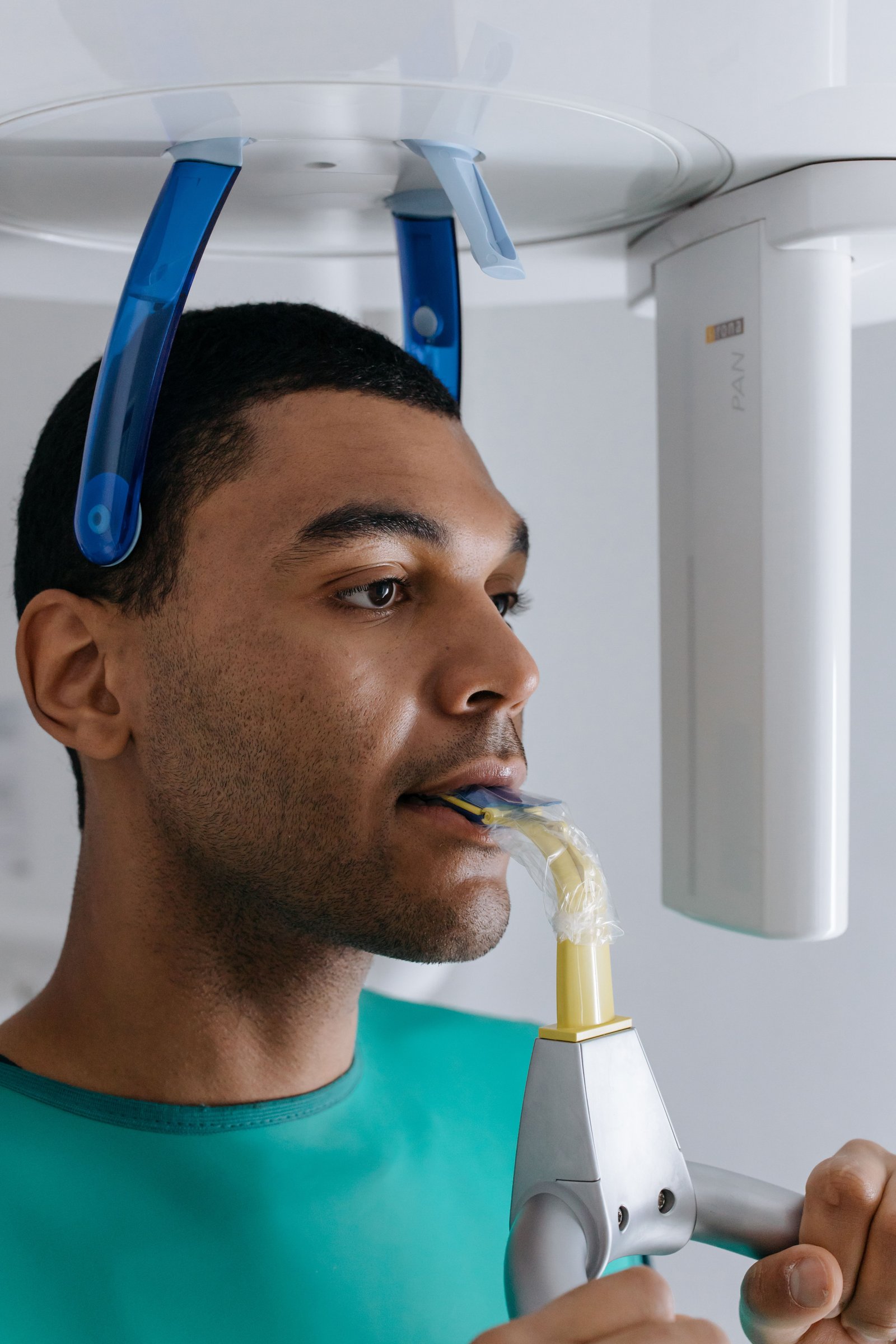
An OPG is a panoramic scanning dental X-ray of the upper and lower jaw. The machine rotates completely around your head in about 15 seconds. It produces a single, flattened 2D image that displays every tooth, the upper and lower jawbones, the maxillary sinuses, and the Temporomandibular Joints (TMJ). It is an essential broad-spectrum screening tool, particularly for assessing impacted wisdom teeth and overall bone health.
3. The 3D Revolution: CBCT Scans
While RVGs and OPGs are excellent tools, they share one fundamental flaw: they compress a three-dimensional human jaw into a flat two-dimensional image. This causes structures to overlap, masking critical anatomical details.
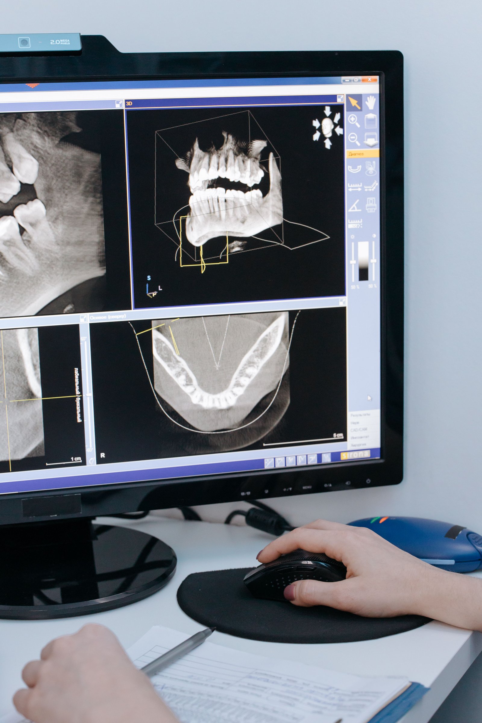
CBCT (Cone Beam Computed Tomography) has revolutionized dental surgery. The scanner rotates around the patient, capturing hundreds of individual images from different angles. A powerful computer compiles these into a highly accurate 3D model that the doctor can rotate, slice, and measure in three dimensions (height, length, and most importantly, width).
Why is CBCT Mandatory for Implants?
When placing a titanium dental implant, the surgeon must know exactly how thick the jawbone is. A 2D X-ray shows height, but not thickness. If a surgeon drills into a bone that is too narrow, the implant will fail, or worse, they may hit the inferior alveolar nerve (causing permanent lip numbness) or perforate the maxillary sinus.
CBCT allows the surgeon to digitally "place" the implant on the 3D computer model before the actual surgery even begins, guaranteeing absolute surgical safety and precision.
4. Intraoral Cameras: Seeing is Believing
We believe that patient education is the cornerstone of ethical dentistry. It is difficult for a patient to understand they have a fractured filling or early decay when it is hidden in the very back of their mouth.
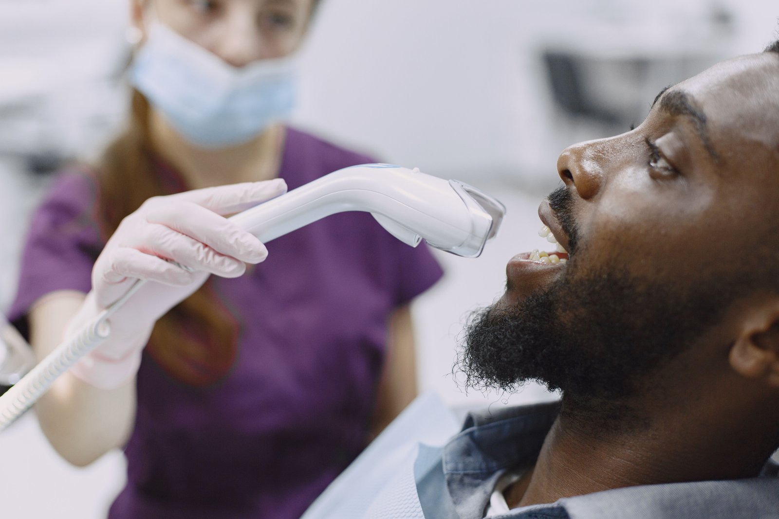
An Intraoral Camera is a highly advanced optical wand, about the size of a dental mirror, equipped with its own LED lighting. The dentist maneuvers it inside the mouth to project live, highly magnified, high-definition video directly onto a monitor in front of the patient. This allows you to see exactly what the dentist sees—plaque buildup, cracked enamel, or receding gums—enabling you to make fully informed, confident decisions about your treatment plan.
5. Radiation Safety: Debunking the Myths
A common concern among patients is the fear of radiation exposure from dental X-rays. In modern dentistry, this fear is largely unfounded.
The ALARA Principle
We strictly adhere to the global medical standard known as ALARA: "As Low As Reasonably Achievable." We only prescribe X-rays when diagnostically necessary, never routinely or unnecessarily. Furthermore, patients are always draped in a heavy lead apron with a thyroid collar to physically block any stray scattered radiation.
Putting Radiation in Perspective
We are exposed to background "cosmic" radiation every day just by walking outside. A single digital dental RVG X-ray exposes you to roughly $0.005$ mSv of radiation. To put that in perspective, that is the exact same amount of background radiation you absorb naturally in just one day of normal life, or during a short 2-hour airplane flight.
6. Advanced Diagnostics in Varanasi
Accurate diagnosis is the foundation of successful medical treatment. Located strategically on BHU Trauma Centre Road in Lanka, Varanasi (easily accessible from Assi Ghat, Samneghat, and Ravidas Gate), Ayaansh Dental Clinic is equipped with modern digital imaging technology.
Directed by Dr. N Srivastava, an MDS Prosthodontist and Implantologist from Banaras Hindu University (BHU), our clinic refuses to operate blindly. Whether planning a complex full mouth rehabilitation or investigating persistent jaw pain, we utilize precise radiographic mapping to ensure your treatments are safe, predictable, and flawlessly executed.
7. Frequently Asked Questions
Yes, routine digital dental X-rays are considered safe during pregnancy. The radiation dose is incredibly low and focused entirely on the head. To guarantee absolute safety for the developing baby, we always drape the pregnant patient in a heavy lead apron to physically block any scattered radiation from reaching the abdomen.
A standard 2D X-ray only shows bone height and length. A 3D CBCT scan provides the crucial third dimension: bone width. It allows the surgeon to see exactly how much bone is available in 360 degrees and digitally map the implant placement to avoid hitting major nerves or sinus cavities.
This depends entirely on your clinical risk factors. If you are highly prone to cavities or have active gum disease, we may recommend "bite-wing" X-rays every 6 to 12 months. If you have excellent oral hygiene and no history of decay, we may only take them once every 2 to 3 years as a baseline check.
Dental X-rays are generally not used to detect soft-tissue oral cancers (like white patches on the tongue or cheek). However, panoramic X-rays (OPGs) and CBCT scans are highly effective at detecting malignant tumors or cysts that have invaded and begun destroying the underlying jawbone.

Dr. N Srivastava
AuthorBDS, MDS (BHU) • Prosthodontist & Implantologist
An expert trained at the prestigious Banaras Hindu University (BHU), Dr. N Srivastava specializes in advanced diagnostics, complex dental implant surgery, and full mouth rehabilitations. She emphasizes precision imaging and uncompromising radiation safety to deliver premium care in Varanasi.